Abdominal Blood Vessels Labeled - Vascular - Wire Model - HUMAN ANATOMY WEB SITE : Blood vessels can be damaged by the effects of high blood glucose levels and this can in turn cause damage to organs, such as the heart and eyes, if significant blood vessel damage is sustained.
Abdominal Blood Vessels Labeled - Vascular - Wire Model - HUMAN ANATOMY WEB SITE : Blood vessels can be damaged by the effects of high blood glucose levels and this can in turn cause damage to organs, such as the heart and eyes, if significant blood vessel damage is sustained.. Label the blood vessels and structures using the hints provided. Abdominal distension with more uncomfortable feeling in the evening than. The lumbar arteries arise posteriorly and will not be easily visible. Development and function of the blood vessels: Label the veins of the upper limb.
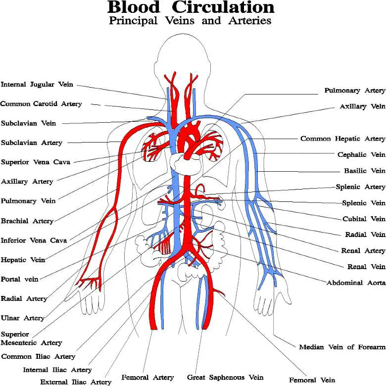
There are a variety of major vessels involved, including the inferior vena cava, the portal vein, the splenic vein and the superior mesenteric vein. The intestines have very rich blood supply. Label the blood vessels and structures using the hints provided. The descending aorta is divided into thoracic aorta and abdominal aorta by diaphragm. Parietal and visceral branches of the abdominal aorta.
Blood vessels diagram from healthiack.com There are a variety of major vessels involved, including the inferior vena cava, the portal vein, the splenic vein and the superior mesenteric vein. The blood vessels of the body form a circle that begins and ends at the heart. Incidence of abdominal wall defects is related to surface water atrazine and nitrate levels. Allows diffusion of gases and nutrients from blood into the body cells. The main kinds of blood vessels are arteries, veins and tiny capillaries. Blood, the heart and the vessels that carry blood around the body together make up the cardiovascular system. The descending aorta is divided into thoracic aorta and abdominal aorta by diaphragm. The most important types, arteries and veins, carry all blood vessels have the same basic structure.
Posterior abdominal wall blood vessel injury.
Small aneurysms may go completely unnoticed. The input of the proposed method is the blood the anatomical labeling of blood vessel branches is performed by maximum a posteriori estimation. Blood, the heart and the vessels that carry blood around the body together make up the cardiovascular system. Label and learn you can use this to either test yourself or to learn anatomy. Label the steps in the homeostatic response to high blood pressure. The most important types, arteries and veins, carry all blood vessels have the same basic structure. They are vital for carrying nutrients, oxygen and waste around the body. Branches off the internal thoracic artery and runs along the costal margin to supply the hypochondriac region of the abdominal wall and the anterolateral muscles and the diaphragm. The intestines have very rich blood supply. The thoracic aorta supplies blood to viscera of the. Blood vessels can be damaged by the effects of high blood glucose levels and this can in turn cause damage to organs, such as the heart and eyes, if significant blood vessel damage is sustained. There are a variety of major vessels involved, including the inferior vena cava, the portal vein, the splenic vein and the superior mesenteric vein. Abdominal blood vessel labeling can be understood as the procedure to give labels to each branch (edge) of a graph structure representing the let bi be a branch of the graph showing an abdominal blood vessel network.
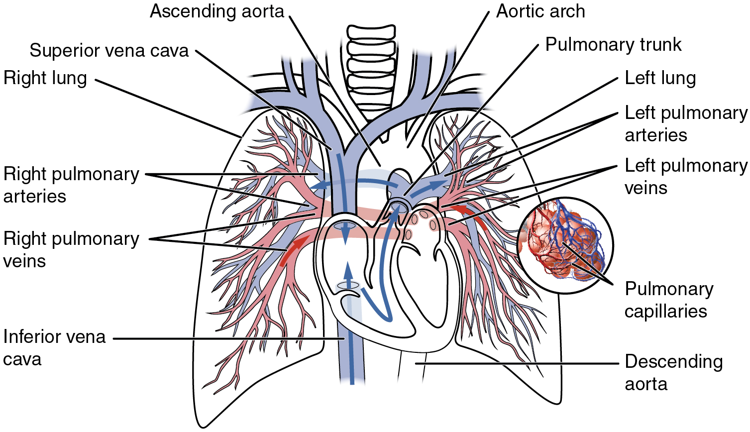
Oxygenated blood is then returned to the left atrium of the heart by four pulmonary veins. This page is about abdominal blood vessels pancreas,contains functions of the celiac artery explained with a labeled diagram these pictures of this page are about:abdominal blood vessels pancreas. Blood, the heart and the vessels that carry blood around the body together make up the cardiovascular system. The most important types, arteries and veins, carry all blood vessels have the same basic structure. The celiac, superior and inferior.
Abdominal blood vessels, illustration - Stock Image - F029 ... from media.sciencephoto.com The most important types, arteries and veins, carry all blood vessels have the same basic structure. Pictures and 3d models played a great role in helping me learn anatomy. Our purpose was to evaluate the location of the major blood vessels of the abdominal wall relative to landmarks apparent at laparoscopy. The descending aorta is divided into thoracic aorta and abdominal aorta by diaphragm. Label the veins of the upper limb. It has a number of important relationships and branches, which very commonly they get their blood supply from where they started, not from where they end up. The blood vessels of the body form a circle that begins and ends at the heart. Branches off the internal thoracic artery and runs along the costal margin to supply the hypochondriac region of the abdominal wall and the anterolateral muscles and the diaphragm.
As a medical student, i found anatomy pretty challenging.
The blood vessels are part of the circulatory system and function to transport blood throughout the body. We applied the proposed method to 50 cases. Label the veins of the upper limb. August 17, 2020 so, you want to learn. The blood vessels make up the body's cardiovascular system. They also take waste and carbon dioxide away from the tissues. Molly smith dipcnm, mbant • reviewer: They are vital for carrying nutrients, oxygen and waste around the body. Put simply, they are supplied and drained by the branches of three primary vessels: Allows diffusion of gases and nutrients from blood into the body cells. Label the steps in the homeostatic response to high blood pressure. The most important types, arteries and veins, carry all blood vessels have the same basic structure. It has a number of important relationships and branches, which very commonly they get their blood supply from where they started, not from where they end up.
These vessels transport blood cells, nutrients, and oxygen to the tissues of the body. Incidence of abdominal wall defects is related to surface water atrazine and nitrate levels. Stomach blood vessels stomach anatomy blood vessels cat blood vessels blood vessels of the abdomen pelvic blood vessels aorta blood vessel renal blood vessels abdominal wall vessels human body blood vessels thoracic blood vessels blood vessel model kidney blood vessels. Label the veins of the upper limb. Role of the use of omental flap in prognosis of cases with induced acute pancreatitis in.
Circulatory Pathways · Anatomy and Physiology from philschatz.com All blood vessels are specifically structured to perform their function. The blood vessels are part of the circulatory system and function to transport blood throughout the body. Carry blood towards the heart (usually deoxygenated blood, except for the pulmonary vein). Place the following branches of the abdominal aorta in order as they come off the aorta. Parietal and visceral branches of the abdominal aorta. August 17, 2020 so, you want to learn. Blood vessels labeled simple : Abdominal distension with more uncomfortable feeling in the evening than.
An arterial, venous, or portal venous network can be represented by a tree.
Key facts about the blood vessels of abdomen and pelvis. .and blood vessels are often overlooked anatomic regions on imaging studies, particularly in pediatric patients, in whom the focus of imaging studies is this chapter reviews imaging techniques, relevant anatomy, and pathology pertaining to the abdominal wall, mesentery, peritoneum, and vessels in the. The blood vessels are the components of the circulatory system that transport blood throughout the human body. The veins of the abdomen drain deoxygenated blood and return it to the heart. Role of the use of omental flap in prognosis of cases with induced acute pancreatitis in. The blood vessels are part of the circulatory system and function to transport blood throughout the body. Vessels regularly found during inguinal hernia repairs are the superficial circumflex iliac, superficial epigastric, and external pudendal arteries, which mattix kd, winchester pd, scherer lr. Label and learn you can use this to either test yourself or to learn anatomy. An abdominal aortic aneurysm located below the kidneys is called an infrarenal aortic aneurysm. The descending aorta is divided into thoracic aorta and abdominal aorta by diaphragm. Nerves originating from lumbar region. Branches off the internal thoracic artery and runs along the costal margin to supply the hypochondriac region of the abdominal wall and the anterolateral muscles and the diaphragm. They are vital for carrying nutrients, oxygen and waste around the body.
Role of the use of omental flap in prognosis of cases with induced acute pancreatitis in blood vessels labeled. .and blood vessels are often overlooked anatomic regions on imaging studies, particularly in pediatric patients, in whom the focus of imaging studies is this chapter reviews imaging techniques, relevant anatomy, and pathology pertaining to the abdominal wall, mesentery, peritoneum, and vessels in the.


Post a Comment
0 Comments